His First Seizure at 36 Was a Brain Tumour in Disguise
Left Frontal Parasagittal Meningioma: How a Single Seizure Led to Diagnosis and Curative Surgery in Thane
By Dr. Bharat Shinde | Neurosurgeon in Thane | neurospinethane.com | Stellars Hospital, Thane
One Seizure. One MRI. A Brain Tumour Found Before It Could Cause Permanent Harm.
A 36-year-old man lost consciousness without warning. His body convulsed. He came around in the emergency department with no memory of what had happened.
His family assumed it was stress. Exhaustion. Perhaps low blood sugar. At 36, with no prior medical history, a seizure seemed impossible to attribute to anything serious.
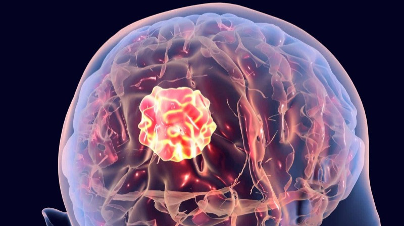
The MRI Brain performed after stabilisation revealed the truth: a left frontal parasagittal meningioma a slow-growing brain tumour that had been silently enlarging for years, pressing on the motor cortex, and had finally announced its presence through a generalised seizure.
He underwent craniotomy with complete excision of the meningioma. He recovered without any neurological weakness. He was discharged in a stable condition.
A first-time seizure is never just a seizure. In this case, it was the only warning sign of a brain tumour growing in a 36-year-old man and the seizure that brought him to hospital was the seizure that saved his life.
| 36
Year Old Male First Seizure Ever |
100%
Meningioma Completely Excised |
0
Neurological Weakness After Surgery |
What Is a Meningioma? The Most Common Benign Brain Tumour
A meningioma is a tumour that arises from the meninges the three-layered membrane that surrounds and protects the brain and spinal cord. Meningiomas are the most common primary intracranial tumours in adults, accounting for approximately 36–40% of all brain tumours.
The overwhelming majority of meningiomas are benign (WHO Grade 1) slow-growing, non-invasive, and curable with complete surgical excision. They do not spread to other organs.
Key facts about meningiomas:
- Most common benign brain tumour accounts for ~36% of all intracranial tumours
- More common in women; peak incidence 40–70 years, but can occur at any age
- Grow slowly often present only when they reach a size sufficient to irritate or compress the brain
- Parasagittal location (adjacent to the superior sagittal sinus at the midline) is one of the most common sites
- Frontal lobe meningiomas often present with seizures, personality change, or headache
- Complete surgical excision (Simpson Grade I or II) is the gold-standard treatment and is curative in most cases
| Parasagittal Meningioma: Why Location Matters |
| A left frontal parasagittal meningioma sits adjacent to the superior sagittal sinus the major venous drainage channel running along the brain’s midline. Surgery in this region requires careful technique to avoid injuring the sinus (which could cause major bleeding or venous stroke) and to protect the adjacent motor strip, which controls movement of the leg on the opposite side. Complete excision in experienced hands is curative. |
His Case: A Tumour Growing Silently for Years, Announced by a Single Seizure
This 36-year-old man had no prior neurological history. No headaches. No weakness. No personality change. Nothing to suggest a brain tumour was growing inside his skull.
That is the nature of frontal meningiomas. The frontal lobe is the brain’s ‘silent area’ in neurosurgical terms a meningioma can grow to significant size in the frontal region before producing obvious symptoms, because the frontal lobe has considerable functional reserve. The tumour gradually displaces rather than destroys brain tissue, and the brain compensates remarkably well until a threshold is crossed.
In this patient’s case, the threshold was a seizure. The tumour had grown large enough to irritate the motor cortex of the adjacent parasagittal region, triggering an abnormal electrical discharge that generalised into a tonic-clonic seizure.
Seizure as the first presentation of a brain tumour is common. Up to 30–50% of brain tumour patients present with a first seizure. This is particularly true for meningiomas adjacent to the cortex, where direct cortical irritation triggers epileptic discharges.
The MRI: Left Frontal Parasagittal Meningioma Identified
Following stabilisation in the emergency department, MRI Brain with gadolinium contrast was performed. The findings were clear:
- A well-defined, homogeneously enhancing extra-axial mass at the left frontal parasagittal region
- The tumour was attached to the dura at the falx cerebri (the midline fold of dura) classic appearance of a parasagittal meningioma
- The adjacent superior sagittal sinus appeared patent no tumour invasion of the sinus
- Mass effect with displacement of the overlying motor cortex laterally
- No surrounding oedema significant enough to require emergency decompression
- Classical MRI features: ‘dural tail sign’ present a hallmark of meningioma
The diagnosis was a left frontal parasagittal meningioma. Given the size, location, and the fact that it had already caused a seizure, surgical removal was clearly indicated.
| Why Surgery Was Necessary – Not Optional |
| A meningioma that has caused a first seizure has reached the threshold of clinical significance. It will not regress spontaneously. It will continue to grow. Future seizures are likely, and the risk of motor weakness, headaches, and cognitive change increases with each passing month of delay. Complete surgical excision at this stage offers the best chance of cure and seizure freedom. |
The Surgery: Craniotomy with Complete Excision of the Meningioma
Following detailed counselling of the patient regarding diagnosis, surgical risks, and the consequences of leaving the tumour untreated, he agreed to and underwent craniotomy with complete excision of the left frontal parasagittal meningioma.
Step 1 – Surgical Planning
Pre-operative MRI venography confirmed the relationship of the tumour to the superior sagittal sinus. Neuronavigation was used intraoperatively to precisely map the tumour boundaries relative to the motor cortex.
Step 2 – Craniotomy Over the Left Frontal Parasagittal Region
A craniotomy was performed over the left frontal parasagittal region, designed to expose the full extent of the tumour and its dural attachment while staying safely lateral to the superior sagittal sinus.
Step 3 – Devascularisation and Tumour Excision
The tumour’s blood supply was systematically coagulated and divided. The meningioma was then progressively debulked from the inside and dissected away from the surrounding brain tissue. The dural attachment at the falx was excised and coagulated (Simpson Grade I excision).
Step 4 – Motor Cortex Protected Throughout
Intraoperative neurophysiological monitoring was used throughout the case to continuously assess the motor cortex and corticospinal tracts. No deterioration in motor signals was recorded during surgery.
Surgical outcome: Meningioma completely excised. Motor function preserved. No new neurological deficits. Patient discharged in stable condition.
Outcome: Seizure-Free, Tumour-Free, Back to Life at 36
- Recovery from craniotomy was smooth and uneventful
- No new neurological weakness motor function in both legs fully intact
- Post-operative MRI confirmed complete tumour excision with no residual lesion
- Anti-seizure medication continued post-operatively no further seizures
- Histopathology confirmed WHO Grade 1 meningioma benign, low recurrence risk
- Patient discharged in stable condition and returned to normal life
At 36, with his entire life ahead of him, he left hospital with a completely excised brain tumour, intact neurological function, and the knowledge that a single seizure investigated promptly had been the event that saved his life.
What Would Have Happened Without Timely Diagnosis and Surgery?
1. Progressive Motor Weakness (Leg Paralysis)
A left frontal parasagittal meningioma grows toward and then onto the motor strip controlling the right leg. Progressive tumour growth would have caused increasingly severe right leg weakness, ultimately leading to spastic paralysis of the lower limb.
2. Refractory Epilepsy
Untreated tumour-related seizures escalate in frequency. Drug-resistant epilepsy is a common consequence of untreated cortical tumours, and the quality of life impact is severe. Early tumour removal gives the best chance of seizure freedom.
3. Cognitive and Personality Change
As the frontal lobe meningioma enlarges, it compresses the frontal lobes bilaterally, causing personality change, poor judgment, memory difficulty, and disinhibition symptoms that families often attribute to ‘stress’ or ‘ageing’ for months before the true cause is found.
Do Not Ignore These Symptoms – They May Indicate a Brain Tumour
- A first seizure at any age this is a brain MRI emergency
- Progressive headaches that are worst in the morning or after lying down
- Subtle changes in personality, behaviour, or memory
- Progressive weakness in one arm or leg without a clear musculoskeletal cause
- Vision changes blurring, double vision, field loss
- Difficulty with speech or word-finding
Brain Tumour Surgery in Thane: Meningioma Excision at Stellars Hospital
Complete surgical excision of meningiomas including complex parasagittal and skull base locations is performed by Dr. Bharat Shinde at Stellars Hospital, Thane. Patients from across the Mumbai Metropolitan Region receive world-class neurosurgical care close to home.
- Neuronavigation-guided meningioma excision
- Intraoperative neurophysiological monitoring for motor and language protection
- Skull base meningioma surgery
- Post-operative MRI and long-term follow-up
Frequently Asked Questions
| Q: Is a meningioma cancer?
The vast majority of meningiomas (>90%) are WHO Grade 1 benign tumours. They do not invade surrounding brain tissue and do not spread to other organs. A small proportion are WHO Grade 2 (atypical) or Grade 3 (anaplastic/malignant), which carry higher recurrence risk and may require radiation after surgery. |
| Q: Will the meningioma come back after surgery?
After complete excision (Simpson Grade I including removal of the involved dura), recurrence risk for WHO Grade 1 meningioma is low (approximately 7–12% over 10 years). Regular MRI follow-up at 1, 3, and 5 years is standard. Incomplete excision carries higher recurrence risk. |
| Q: Can a meningioma be observed without surgery?
Small incidental meningiomas without symptoms or oedema can be watched with regular MRI. However, a meningioma that has caused a seizure, is causing oedema, or is adjacent to critical structures (motor cortex, sinus) warrants surgical removal to prevent progressive neurological decline. |
| Q: How long is recovery after meningioma surgery?
Most patients are discharged within 4-6 days after craniotomy. Return to desk work occurs within 4–6 weeks. Anti-seizure medication is typically maintained for 1–2 years post-operatively, with gradual weaning if the patient remains seizure-free. |
| Q: Is meningioma surgery available in Thane?
Yes. Dr. Bharat Shinde at NeuroSpine Thane and Stellars Hospital performs craniotomy for meningioma excision including complex parasagittal and convexity meningiomas. Patients from Thane, Navi Mumbai, Kalyan, and across the MMR are treated here. |
| Dr. Bharat Shinde | Neurosurgeon in Thane | Stellars Hospital
A first seizure needs an MRI today, not next week. NeuroSpine Thane at Stellars Hospital provides emergency neurosurgical evaluation and brain tumour surgery for patients across Thane and the Mumbai Metropolitan Region. Serving: Thane West, Thane East, Mulund, Airoli, Belapur, Kalyan, Dombivli, Bhiwandi, Mira-Bhayandar, Vasai-Virar, Navi Mumbai and all of MMR neurospinethane.com | Stellars Hospital, Thane |